

Patented & innovative electrophysiology diagnostic catheter
EP Bioptom is an innovative electrophysiology diagnostic catheter that combines two critical functionalities:
 the ability to perform biopsy
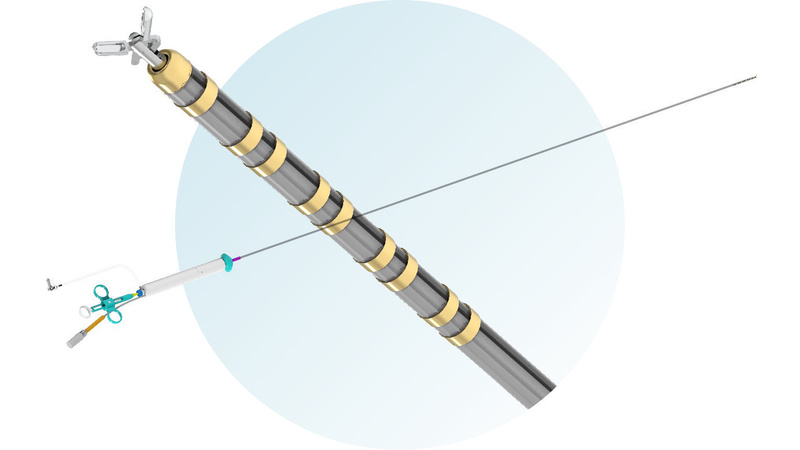
Structure of EP Bioptom
The design of EP Bioptom includes biopsy forceps and a catheter with diagnostic rings enabling monitoring using a 3D electroanatomical mapping system (EAM, 3D-EAM). Additionally, the EP Bioptom catheter can be connected to an irrigation system, allowing the internal channel of the catheter to be flushed with saline solution.

About the heart biopsy
Cardiac biopsy, also known as endomyocardial biopsy (EMB), is an invasive procedure that provides morphological, immunohistological and structural examination. Myocardial biopsy is performed in specific heart diseases such as unexplained congestive heart failure, sarcoidosis, amyloidosis, or storage diseases.
An indication for biopsy is also a suspicion of neoplastic processes, giant-cell myocarditis, not responding to treatment and idiopathic acute heart failure, heart failure in individuals with eosinophilia, arrhythmia, suspicion of arrhythmogenic cardiomyopathy, conduction disorders and necessity of transplanted heart examination for possible rejection.
Biopsy procedure
During EMB 3 to 10 samples of the right and/or left ventricle are taken. EMB is performed using special surgical instruments - biopsy forceps (also called bioptome), which are inserted into the heart using dedicated catheters.
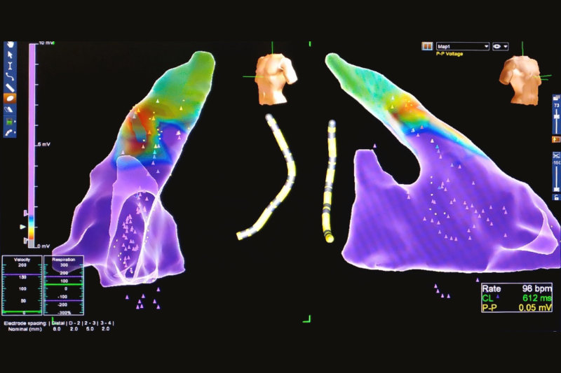
Mapping system
Electroanatomical mapping systems allow to create three-dimensional potential map to identify areas of the heart with pathological electrical function. The electrophysiologist performing the potential maps has the ability to map the area of the border zone, scar and healthy heart muscle. In addition, 3D mapping allows for the determination of the conduction system and valve area, which reduces the risk of biopsy from the conduction system/valve area and the related complications.

Application of technology
Proper use of this technology allows precise localization of the source of the arrhythmia, determination of the heart chamber 3D-geometry, anatomically interesting areas, catheter manipulation and positioning without the need for using X-rays, which are currently used during every EMB procedure. 3D-EAM systems facilitate the navigation in the heart and increase the procedure success, especially for complex cardiac arrhythmias and unusual cardiac anatomy.
A new dimension of security
The ability to map the heart with the EP Bioptom catheter offers new possibilities in the field of invasive cardiac surgery, providing a much safer solution than previously used methods.